Dental X-ray Fears: Concerns Over Dental Radiation Exposure Unfounded
By Dr. Paul Wilke, DDS, June 20, 2013
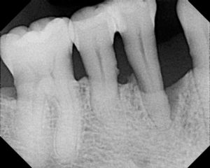
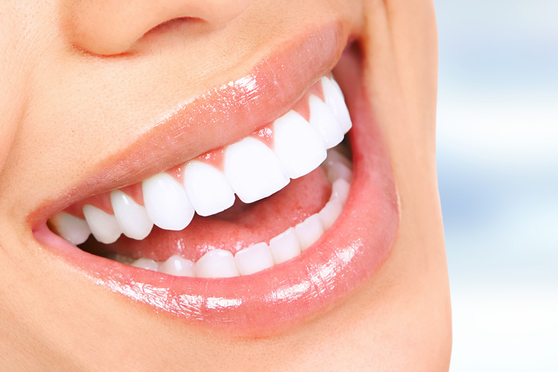
X-ray images are a dentist’s most valuable diagnostic tool. They are much more important than even an oral examination. I can’t provide the highest quality of care to my patients without them. X-rays are routinely used to detect decay between teeth and under fillings and crowns. Most of the time, this decay cannot be seen in the mouth visually. X-rays are also used to find infected, abscessed teeth and to determine bone levels in periodontal disease. These images are absolutely necessary before extractions to see root anatomy, sinus levels, and inferior alveolar nerve positions. In addition, they are necessary in children to find congenitally missing teeth and show proper eruption patterns to determine orthodontic requirements.
Unfounded Fears
In March of 2012, an article in the journal Cancer tried to link dental X-rays with an increased risk of developing meningioma, a non-malignant brain tumor. The article was picked up by mainstream media because of the “fear factor,” and the sensationalized reporting of the article resulted in many patients feeling fearful of receiving dental X-rays. In my view, the original article fit the definition of “junk science” and the resulting media reporting created a lot of needless fear.
A major flaw with the original study was its use of anecdotal evidence. The study asked patients to recall from memory their dental X-ray history – for some study participants this meant recalling dental visits going back more than 70 years. Research should be based on facts and not memories of what happened 75 years ago. Also, the dental X-ray exposure of many of these patients was back in the 1950s and 1960s when X-ray equipment, film and techniques were very different. Modern digital X-ray sensors, as used by many dentists today, along with the advanced imaging software and newer generation X-ray units, have reduced radiation to As Low as Reasonably Achievable (ALARA).
One of the outcomes of the study showed the odds ratio risk from bitewing X-rays to be 1.2-2.0 and the ratio from a full mouth series to be 1.0-1.2. This is biologically impossible unless X-rays reduce the incidence of tumors! A full mouth series of radiographs involves taking 18 intraoral films, including two to four bitewings. It obviously makes no sense that two to four films could cause more damage than 18 films. The study also failed to take into account participants’ medical X-ray history. This is a significant omission when you consider that head CT exposures, for example, have 15 times more equivalent dose than dental radiographs. Previous research efforts to link head CT exposures, skull X-rays, or sinus radiographs to meningiomas have all been unsuccessful according to a Medscape article (www.medscape.com). Decades of study involving Japanese atomic bomb survivors as well as other exposed populations have not reliably demonstrated evidence of increased cancer risk below 100 mSv (millisievert) radiation, a huge dose.
Everyday Radiation Exposure
Radiation is around us every day. We cannot escape it. The average person on earth is exposed to 3.6 mSv of radiation per year from background sources. This comes from outer space, the earth, natural materials including foods, and even other people. As a general rule, this background radiation doubles with each one mile of elevation change. Therefore, people living in Leadville, Colo., at 10,100 feet will have considerably more background radiation than those of us in San Antonio at about 700 feet.
Dental X-ray Risk: Minimal
Equivalent dose is the term used to measure the amount of radiation a body part is exposed to. There are many terms but a unit of equivalent dose in the U.S. is called a Rem. Another term used outside the U.S. is called the Sievert. 100 Rems = 1 Sievert. Exposure to medical radiation is generally measured in millirems (mRem) and millisieverts (mSv) which is 1/1000 as much.
Since each of us receives 3.6 mSv of radiation per year from background radiation, we can divide this by the amount of radiation in one digital bitewing X-ray which is about .00325 mSv and come up with an equivalent number of bitewing X-rays per year: 1,108. Therefore, each person is already receiving the equivalent of 1,108 bitewing X-rays per year (about three bitewings per day) through background radiation. My patients generally receive two to six bitewing X-rays each year – that’s all. The risk of missing easily diagnosable dental diseases that can be treated with simple procedures far outweighs the small exposure to radiation, especially with digital radiography.
In my practice, I have seen hundreds of teeth needlessly lost due to infrequent dental exams. If we had found the problem sooner those pulled teeth would still be in my patients’ mouths. Dental X-rays are absolutely the safest type of medical X-ray procedures performed, so don’t let an unfounded fear of dental radiation stop you from getting the best preventive and diagnostic dental care available.
By Dr. Paul Wilke, DDS